
In brief
- Shoulder dislocation corresponds to the complete dislocation of the head of the humerus out of the glenoid of the scapula.
- In Children, instability related to hyperlaxity can explain dislocations in several directions. In Adolescents, the mechanism is more often a sports trauma.
- A sudden pain, an arm held immobile, and a visible deformation require an emergency evaluation.
- Treatments combine medical repositioning, Immobilization, Rehabilitation, and, in some recurrences, Surgery.
Understanding shoulder dislocation in children and adolescents
The shoulder joins the head of the humerus, the long bone of the arm, and a shallow cavity of the scapula called the glenoid. This joint favors the range of motion. It allows throwing, climbing, catching, swimming, or putting on a garment, but this great mobility makes it less stable than a joint like the hip.
During a dislocation, the head of the humerus comes completely out of the glenoid. A subluxation corresponds to an incomplete displacement. The joint sometimes returns to place on its own, but the episode should not be trivialized, especially if it happens several times. Ligaments, the joint capsule, tendons, and sometimes cartilage structures can be stretched or injured.
A truly dislocated shoulder usually causes immediate pain and an inability to use the arm as before the accident. The child or adolescent often holds their limb against their body, as the slightest movement increases the pain. An asymmetry may be visible in the shoulder contour, with an unusual bony prominence.
The frequency and mechanism vary significantly with age. Traumatic shoulder dislocation remains uncommon in young children. The bones and supporting tissues do not respond the same way as in adolescence. In younger children, repeated pain, sensations of dislocation, or instability can be due to hyperlaxity. Connective tissues are then more stretchable than average.
From adolescence, especially in young athletes, the scenario is often clearer. A fall on the arm, contact in handball or rugby, a powerful serve in tennis, a poorly controlled landing in gymnastics or skiing can cause the humeral head to dislocate forward. This form is called anterior dislocation.
The terms “dislocated shoulder,” “out of joint,” or “displacement” are used by families to describe several realities. A clavicle fracture, a humerus fracture, a deep bruise, or an acromioclavicular sprain can also give the impression that the shoulder has changed shape. Only a medical examination, most often supplemented by an X-ray, can distinguish these situations.
You can consult this guide on shoulder dislocation in children and adolescents to keep simple reference points before a consultation. The word “dislocation” describes a specific joint problem. It alone does not predict the recovery time, as it depends on age, associated injuries, the sport practiced, and whether there have been previous episodes.
Identifying causes according to age and trauma mechanism
In adolescents, the most frequent cause is a Trauma during sports activity or a fall. The arm is often away from the body, turned outward and projected backward. Orthopedists talk about a cocked position because it resembles the preparatory gesture of a throw. In this position, the head of the humerus can overcome the anterior rim of the glenoid.
Contact sports expose more to direct impacts. Rugby, football, judo, basketball, and handball are involved. Disciplines where the arm frequently passes above the head, such as tennis, volleyball, athletics, climbing, or gymnastics, also strongly solicit the joint. This does not mean these activities are dangerous by principle. Technique, muscle strengthening, fatigue, and history modify the risk.
In younger children, multidirectional instability can manifest without major impact. The shoulder seems to move forward, backward, or downward depending on the movement. Generalized hyperlaxity may be observed when the child shows significant flexibility in several joints, for example elbows, knees, or fingers. This flexibility is not a disease by itself. However, it deserves evaluation when accompanied by pain, repeated dislocations, or difficulties in daily gestures.
Some episodes occur around 8 or 9 years old in very flexible children, sometimes without identifiable fall. The muscular system then does not sufficiently ensure the centering of the humeral head in the glenoid. The rotator cuff muscles and those stabilizing the scapula play a guiding role. When they lack endurance or coordination, the already very extensible capsule is no longer enough to contain extreme movements.
| Observed situation | Common mechanism | Usual outcome |
|---|---|---|
| Adolescent after fall, contact or throw | Traumatic anterior dislocation | Emergency, X-ray, repositioning then rehabilitation |
| Very flexible child with repeated instability | Multidirectional instability linked to hyperlaxity | Specialist assessment and progressive muscle strengthening |
| Pain after impact without deformation | Possible bruise, sprain or fracture | Medical examination depending on pain and mobility |
| Brief sensation of dislocation then immediate return | Possible subluxation | Scheduled or prompt consultation depending on pain and recurrence |
A rarer cause can be sought when hyperlaxity is very marked, familial, or associated with pain in many joints, frequent sprains, or particularly fragile skin. The doctor may then refer to a specialized consultation, sometimes in genetics, to explore collagen diseases. This approach does not concern most flexible children.
The distinction between these two profiles truly guides care. Surgery aiming to block forward instability in a traumatized adolescent does not necessarily meet the needs of a child whose shoulder moves in several directions. The correct treatment therefore starts with a thorough understanding of the cause.
Recognizing symptoms that require quick management
The pain of a dislocated shoulder appears suddenly. It is often very intense from the accident, then increases when the child tries to move the arm. The adolescent sometimes describes a very clear sensation of dislocation, followed by a blockage. Younger children do not always use these words. They may cry, refuse to be touched, or spontaneously keep the elbow close to the torso.
A shoulder deformation associated with intense pain after a fall or impact should be considered an emergency. The normally rounded contour may appear flattened. A bump may be noticeable in front or under the skin. This sign is not systematic, especially if body build or muscle contractions mask the relief modification.
Possible symptoms should be considered together rather than separately.
- The arm remains immobile, supported by the other hand or held against the stomach.
- Pain is sharp and occurs just after an impact, fall, or forced movement.
- The child can no longer raise the arm or turn the shoulder without strong pain.
- A shape difference between the two shoulders or an unusual prominence appears.
- Tingling, numbness, or a cold hand must be reported immediately.
These signs do not allow parents to make a diagnosis at home. They help decide the urgency level. An X-ray is generally performed in the emergency room before reduction to verify the absence of an associated fracture. In adolescents, a high-energy fall can also cause bone or cartilage injury that will modify the course of management.
Do not attempt to reposition the shoulder at home, even if you have seen a maneuver on a video or if a relative thinks they know the gesture. Inappropriate traction can increase pain, further displace the joint, or worsen an unnoticed fracture. Instead, put the arm in the least painful position, with a cushion or improvised sling, without tightening.
A cold pack wrapped in a cloth can be applied for a few minutes on the painful area, without direct contact with the skin. If the child can take a painkiller usually allowed for them, respect the dosage already indicated by a professional or the one on the prescription. This measure should not delay the trip to the emergency room.
The situation is different when there is neither deformation nor major incapacity, but pain persists after training or a false movement. A medical consultation in the following days then allows evaluation of a sprain, tendinitis, instability, or subluxation. In an adolescent who feels several times that their shoulder “slips,” waiting for a complete dislocation exposes them to new injuries.
Quick evaluation helps relieve pain, rule out a fracture, and protect nerves and vessels near the joint. Once the emergency is recognized, care can proceed much more calmly.
Appropriate treatments between reduction, immobilization, and rehabilitation
In the emergency room, the team first assesses pain, hand sensitivity, circulation in the fingers, and mobility. An X-ray is most often performed. It confirms the position of the humerus and looks for a fracture. In some young athletes, a CT scan or MRI can be proposed later if the specialist suspects damage to the bony rim of the glenoid, cartilage, or ligaments.
Repositioning is called reduction. It consists of gently guiding the humeral head into the glenoid. Muscle relaxation is decisive because pain causes a reflex contraction that makes the movement more difficult. Painkillers are administered and nitrous oxide may be used in some departments to reduce anxiety and facilitate relaxation. More significant sedation is sometimes necessary depending on age, pain intensity, and clinical situation.
Relief is often clear after reduction, but the shoulder remains sensitive. Ligaments and muscles have undergone sudden tension. Pain may persist several days, sometimes longer, with a progressive decrease. Prescribed medications must be given as directed. Pain that intensifies instead of decreasing, fever, or blue, very cold, or numb fingers justify a prompt new medical consultation.
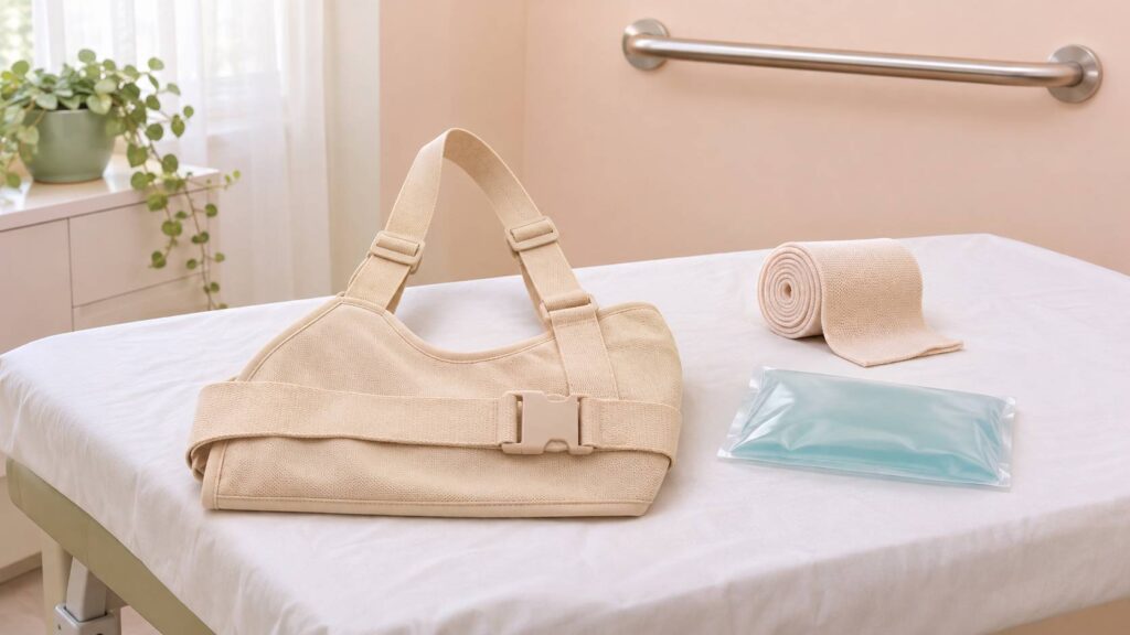
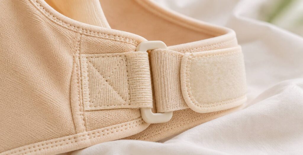
Immobilization with the elbow to the body often lasts about three weeks after a first traumatic dislocation, with a duration adapted by the specialist. The splint protects the joint during the tissue healing period. It does not mean that the child must remain totally inactive. Walking and allowed activities can be maintained, but arm movements, ball games, carries, and risks of falls are temporarily limited.
Rehabilitation starts after the period set by the doctor. It often takes place with two to three weekly sessions, over a duration that can extend up to three months. Recovery is not a simple succession of stretches. The physiotherapist first teaches movements without pain, then works on scapular stability, rotator cuff strength, coordination, and gestures specific to the sport practiced.
In hyperlax children, the strategy changes. Physiotherapy plays a central role and can be extended by strength programs regularly resumed. The goal is not to make a joint rigid. It is to provide muscles with references and enough endurance to control excessive mobility. Breaststroke can sometimes be included in an individualized program, while repetitive movements above the head, such as back crawl, are sometimes avoided depending on clinical examination.
Returning to sport is not decided solely by the disappearance of pain. The shoulder must have regained satisfactory mobility, strength close to the opposite side, and reliable control in fast movements. After simple reduction, the timetable depends on the sport, the presence of lesions, and the specialist’s opinion. Returning too soon to a contact sport increases the risk of a new episode.
When shoulder dislocation surgery may be discussed
Surgery is generally not offered after an isolated first dislocation without major injury, especially if recovery evolves properly with the splint and Rehabilitation. In sporty adolescents, the question can however be discussed in case of recurrences, lesions identified by imaging, or high risk of new dislocation related to the sport practiced. The goal is to protect the joint before repeated episodes further damage the bone, cartilage, or stabilizing tissues.
The orthopedic surgeon considers several factors. The number of accidents counts but is not sufficient alone. Age, type of sport, direction of dislocation, quality of recovery, glenoid lesions, and capsule condition are also analyzed. This decision is prepared after a precise assessment, without wrongly opposing rehabilitation and surgery. These two approaches often complement each other.
In some anterior instabilities, a capsulorrhaphy may be proposed. The intervention aims to tighten the capsulo-ligamentous tissues that have become too loose or damaged. Another technique, the bone block, consists of placing a bone fragment in front of the glenoid to create a barrier to the humeral head’s exit. It can be chosen when there is bone loss or in some athletes very exposed to contacts and shocks.
After surgery, the splint, temporary sports cessation, and rehabilitation remain necessary for several months. A return can be considered between three and six months depending on the procedure performed, the sport, and functional results. The timetable should not be dictated by a close competition. The achieved stability also depends on consolidation and progressive muscle work.
In children with multidirectional instability related to hyperlaxity, surgery is reserved for exceptional situations. Tightening ligaments of a very flexible shoulder can yield an incomplete result or even shift instability to another direction. A block placed in front may not address a problem also involving the back or bottom of the joint. Specialized teams can then discuss the case collegially.
Repeated dislocations can have a broader impact than just physical pain. Some children stop participating in playground games, dread sports class, or monitor every movement of their arm. This caution is not a lack of courage. The brain associates a gesture with intense pain and triggers a protective anticipation. Pain management, with a pediatric pain specialist when it becomes chronic, can include appropriate medication, physiotherapy, sophrology, or psychological support.
You will also find additional explanations on the causes, symptoms, and treatments of this shoulder injury. Regular follow-up helps adjust activities without unnecessarily removing the child or adolescent from sport, school, and daily life.
Can a shoulder dislocation reduce itself?
A subluxation can sometimes reduce spontaneously, but even a brief sensation of dislocation deserves medical advice, especially if it recurs. A true painful dislocation with deformation requires emergency care.
Can we wait until the next day if the pain decreases?
After a fall with deformation, inability to move the arm, tingling, or very severe pain, do not wait. These signs require prompt evaluation to check for dislocation, fracture, or nerve injury.
How long does rehabilitation last after a dislocation?
After a traumatic dislocation, it can last several weeks and sometimes up to three months. The duration depends on age, injuries, the sport practiced, and regained stability.
Should a very flexible child stop sports?
Not necessarily. Activity can be adapted with the doctor and physiotherapist. Muscle strengthening and learning controlled movements are often more helpful than a total and prolonged stop.