

In brief
- Otospongiosis, also called otosclerosis, is a rare bone disorder that mainly hinders the mobility of the stapes, a small bone in the middle ear.
- It most often causes a progressive hearing loss, sometimes accompanied by tinnitus or a sensation of imbalance.
- The otospongiosis diagnosis is based on ENT examination, audiometry, and, depending on the situation, a CT scan of the petrous bones.
- A hearing aid or otospongiosis surgery can significantly improve hearing when stapes fixation is confirmed.
Otospongiosis and middle ear understanding the mechanism of hearing loss
Otospongiosis is a disease of the bone surrounding the inner ear, called the otic capsule. It can also be referred to by the term otosclerosis. These two words describe the same pathology, although professionals may prefer one or the other depending on habit.
To understand what happens, you need to look at the path of sound. Sound vibrations reach the eardrum, then are transmitted by three small bones located in the middle ear: the malleus, incus, and stapes. The stapes is the last in this chain. It transmits vibrations to the inner ear, where they are converted into nerve messages interpreted by the brain.
In otospongiosis, local bone remodeling is disturbed. Areas of bone with different quality appear near the footplate of the stapes, that is, its part in contact with the oval window. The stapes progressively loses its mobility. Vibrations travel less effectively, which creates a conductive hearing loss.
The sound is present around the person, but its mechanical transmission to the inner ear becomes less effective. This distinction explains why some conversations seem audible in a quiet environment, whereas exchanges quickly become tiring in a restaurant, a family gathering, or a noisy place.
This disease does not reflect a lack of attention to hearing or bad lifestyle habits. It corresponds to a specific biological disorder. In France, frequently cited estimates place its prevalence between 0.2% and 0.5% of the population. Therefore, it remains rare, while representing a known cause of acquired hearing troubles in young adults.
The discomfort may affect only one ear at first. The other ear may remain stable for a long time or be affected later. This sometimes asymmetrical progression is confusing, as the person feels that their hearing varies depending on the seat at the table, the side of the telephone, or the direction from which a voice comes.
Otospongiosis is not always limited to mechanical transmission only. When bone lesions are close to the inner ear structures, a sensorineural component may be added. The hearing loss then becomes mixed. This situation requires a more detailed ENT assessment, as the proposed solution depends on the precise type of deficit measured by audiogram.
The difficulty is often invisible to those around. An affected person may respond incorrectly, regularly ask to repeat, increase the volume of the television, or seem less available in group discussions. These behaviors do not signify disinterest. They sometimes reflect the constant effort made to mentally reconstruct incompletely heard sentences.
For a parent, this auditory fatigue can be worsened by night awakenings, sharp cries, rapid exchanges with caregivers, or the many simultaneous sounds of family life. Hearing loss should then be taken seriously without waiting for it to become disabling. The goal is not to be alarmed but to regain easier communication daily.
Origin of otospongiosis and factors that may promote its expression
The origin of otospongiosis is not limited to a single cause. Medical knowledge mainly supports the existence of a genetic predisposition. Family history is found in some affected individuals, with a transmission that can appear irregular from one generation to another.
This variability is notably explained by incomplete penetrance. A person may carry a predisposition without developing marked symptoms. In the same family, one relative may need surgery, while another experiences mild discomfort or never has identified hearing loss.
A family history does not predict a certain progression, but it gives a concrete reason not to trivialize progressively decreasing hearing. When a parent, brother, sister, or grandparent has undergone stapes surgery or used hearing aids relatively young, this information should be reported to the ENT doctor.
Otospongiosis is more frequently diagnosed in women. Hormonal change periods, such as puberty, pregnancy, or menopause, are sometimes associated with onset or an impression of accelerated symptoms. This does not mean pregnancy causes the disease. The link between hormones and progression remains complex, and each situation should be individually assessed.
Pregnancy represents a time when medical vigilance is useful without becoming anxiety-provoking. Tinnitus, a blocked ear, or hearing loss can have several explanations. A change in hearing alone does not allow concluding otospongiosis. High blood pressure, anemia, infection, earwax buildup, or another ENT condition may also be involved.
New tinnitus during pregnancy justifies discussing with the professional following the pregnancy, especially if accompanied by unusual headaches, visual disturbances, sudden swelling of the face or hands, pain under the ribs, or a feeling of malaise. These signs require rapid evaluation of blood pressure and general condition.
Hormonal contraception often raises questions. Commonly prescribed contraceptives are not considered a proven cause of otospongiosis. A person already followed for this condition can nonetheless discuss their contraceptive treatment with their gynecologist and ENT specialist, particularly if a high-dose formulation is contemplated or if hearing seems to change.
Infectious hypotheses, notably around measles, have been studied. They do not explain all cases and do not replace the genetic explanation in familial forms. In 2026, no common infection allows predicting or confirming this diagnosis by itself.
Careful follow-up protects better than anxious surveillance. Noting the onset date of discomfort, the affected side, situations where it occurs, and the presence of tinnitus helps the ENT to rebuild the disorder’s history. This simple observation is more useful than testing one’s hearing by constantly increasing the volume on devices at home.
Additional information on initial markers and signs to have evaluated is available in this guide devoted to the origin and signs of otospongiosis. Dialogue with an ENT remains the right path when discomfort persists, as the examination distinguishes stapes involvement from other common causes of hearing loss.
Otospongiosis symptoms recognizing progressive hearing loss
Otospongiosis symptoms most often begin in adolescence or young adulthood. Forms beginning in childhood exist but are rare. The main sign is a slow decrease in hearing, without sudden pain or fever in most cases.
The person sometimes hears better in some contexts and worse in others. Low voices or quiet sounds become less distinct. Consonants, which give words their precision, can be confused. Sentences then appear less clear, especially if several people speak simultaneously.
Tinnitus is frequent. It can take the form of a whistle, buzzing, whooshing, or continuous noise perceived in one or both ears. Its intensity often varies with fatigue, stress, lack of sleep, or evening silence. Tinnitus does not automatically indicate otospongiosis, but its association with progressive hearing loss justifies scheduling a consultation.
Dizziness or instability sensations can also occur, more rarely. They should not be attributed too quickly to this disease. True vertigo gives the impression that the room is spinning or the body is tilting. It differs from lightheadedness related to fatigue, dehydration, or low blood pressure.
| Observed situation | What it may indicate | Appropriate response |
|---|---|---|
| Slow hearing loss over several weeks or months | Otospongiosis or other cause of progressive deafness | Make an appointment with an ENT for a hearing assessment |
| Blocked ear after swimming or recent cold | Earwax, inflammation, or transient ventilation disorder | Consult depending on duration and avoid manipulations in the canal |
| Sudden hearing loss in one ear | Possible ENT emergency | Seek medical advice the same day |
| Tinnitus with headaches and visual disturbances during pregnancy | Obstetrical or hypertensive cause to investigate | Contact maternity, midwife, or gynecologist promptly |
Sudden hearing loss, even without pain, does not correspond to the usual otospongiosis picture and requires urgent medical evaluation. Waiting several days in this situation can delay care for sudden deafness, which has another cause.
The impact often goes beyond listening alone. The person may avoid noisy meals, feel irritated after a day of conversations, or fear not hearing a child calling from another room. Exhaustion linked to decoding effort is real. It deserves recognition in the medical discussion.
A simple home test does not replace audiometry. Comparing the two ears with headphones, asking a close one to whisper, or using an app neither measures the deficit precisely nor identifies its cause. These trials can however help describe what has changed, provided they do not delay consultation.
Consultation is particularly indicated when discomfort lasts more than a few weeks, becomes asymmetrical, is accompanied by persistent tinnitus, or forces communication habit changes. Talking face to face with the person, reducing background noise during interaction, and announcing one’s presence before speaking from another room can already ease listening load while waiting for assessment.
This approach does not mean medicalizing every distraction or listening difficulty. It helps distinguish a period of fatigue, a often reversible mechanical problem, and an ear disease that requires precise follow-up.
Otospongiosis diagnosis exams that help choose the right response
The otospongiosis diagnosis is made by an ENT specialist. It begins with a detailed discussion about the evolution of hearing difficulties, family history, tinnitus, vertigo episodes, and ongoing treatments. This timeline has real value, as slow progression and sometimes unilateral character guide exams.
Otoscopy allows observing the ear canal and eardrum. In many cases, this exam is normal in otospongiosis. This sometimes surprises, as the perceived discomfort is significant while the eardrum appears intact. The involvement lies behind it, at the level of ossicles and bone capsule.
Audiometry is the central examination. It measures hearing thresholds with sounds transmitted by headphones and bone vibration. It differentiates conductive, sensorineural, or mixed hearing loss. The result is not reduced to a hearing percentage. It indicates which frequencies are affected and how sound reaches the inner ear.
Tympanometry and stapedius reflex testing can complete the workup. They provide information about the middle ear’s mechanical function. Absence or modification of certain reflexes can strengthen suspicion but alone is not sufficient to confirm diagnosis.
CT scan of the petrous bones, performed with a technique adapted to the fine ear structures, helps visualize foci compatible with otospongiosis and prepare for possible surgery. It is not systematic for every hearing discomfort. The ENT requests it when it provides useful information for the decision.
The workup does not only aim to name the hearing loss, it specifies if the stapes is really fixed and if surgery can bring coherent benefit. This precision avoids proposing a standard solution to situations that are not alike.
The follow-up rhythm depends on the discomfort and the exam results. A mildly affected person with stable hearing may benefit from spaced audiograms. If hearing loss worsens, if work becomes difficult, or family exchanges complicate, a more active discussion on compensation options is justified.
Certain signs require a quicker appointment. Sudden deafness, intense and persistent vertigo, facial weakness, bloody ear discharge, severe pain with fever, or head trauma do not correspond to usual simple otospongiosis progression. The attending physician, an emergency department, or an ENT must then guide care depending on context.
The workup benefits from including practical consequences. A person who poorly hears alarms, a child’s calls, work instructions, or phone conversations should not minimize this impact. These elements participate in choosing between surveillance, hearing aids, and surgery.
Those close to the person can support this step without speaking for the affected. Reformulating rather than shouting, keeping the face visible, and limiting a turned-on television during a conversation already change the quality of exchanges. Hearing is not merely a measurement on a graph; it organizes safety and daily fluidity.
Otospongiosis treatment between hearing aid and stapes surgery
Otospongiosis treatment depends on several elements. The severity of hearing loss, its type, the condition of the other ear, CT scan results, the person’s activities, and preferences all matter. There is no pill capable of permanently restoring the mobility of an ankylosed stapes.
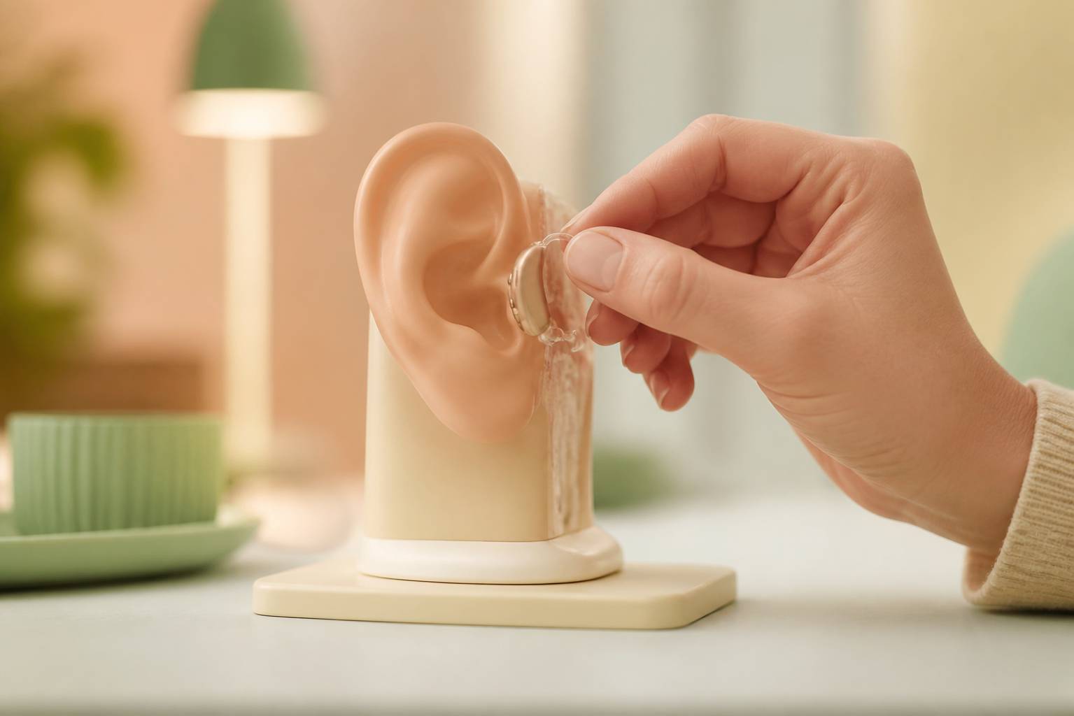
A hearing aid is an effective option for many people. It amplifies sounds to compensate for transmission loss. Current devices can be discreet and adjusted according to the audiogram. An adaptation period is usual, as the brain must relearn to process sounds that have become less present.
Use of a hearing aid does not mean the situation is final or that surgery becomes impossible. It can be chosen because it matches the person’s wishes, because the ear does not present favorable conditions for surgery, or because the deficit associates several mechanisms. A hearing care professional works in conjunction with the ENT to adjust settings and assess real comfort.
Otospongiosis surgery, often called stapedotomy or stapedectomy, aims to bypass stapes fixation. The surgeon creates a small opening in the footplate and places a prosthesis linking the incus to the inner ear. Older descriptions involved removing the stapes. Current techniques most often seek to preserve the existing structures as much as possible.
Surgery seeks to restore vibration transmission; it does not guarantee perfect hearing in all situations. The expected benefit depends notably on the proportion of conductive hearing loss. When the inner ear is also affected, the gain can be more limited, and hearing aids may remain useful.
The intervention is proposed after detailed discussion with an ENT surgeon experienced in this practice. Results are often favorable on the mechanical component of hearing loss, but no procedure is without risks. Transient vertigo, taste alteration, eardrum perforation, worsening hearing, or more severe deafness are rare but must be explained before decision.
After surgery, improvement is not always immediate. The ear needs to heal and hearing stabilizes progressively. Instructions are given concerning water in the ear, physical efforts, airplane travel, or activities exposing to significant pressure changes. These vary with technique and surgeon.
Monitoring the other ear remains useful, as the condition can be bilateral. This surveillance does not mean a new operation will be necessary. It helps detect evolution on time before communication difficulties become overwhelming.
Pharmacological approaches have been studied in the past, notably around sodium fluoride or treatments acting on bone metabolism. They do not constitute a standard treatment for stapes ankylosis. Hormone replacement therapy should not be prescribed to treat otospongiosis without a specific medical indication.
Additional information on follow-up choices and available solutions can be found in this resource on otospongiosis signs. The best decision is the one that takes into account examinations, lived impact, and the role the person wishes each option to have in daily life.
Can otospongiosis affect both ears?
Yes. It can start on one side and then affect the other ear later. The progression is very variable, which justifies regular audiometric follow-up with an ENT doctor.
Can sudden hearing loss be due to otospongiosis?
This is not the usual picture. Otospongiosis most often causes progressive hearing loss. Sudden deafness in one ear requires urgent medical consultation the same day.
Does pregnancy cause otospongiosis?
Pregnancy alone does not cause this disease. Hearing changes may be noticed during this period, but they require evaluation as several causes are possible.
Does the hearing aid prevent later surgery?
No. Hearing aids can be a long-term or temporary solution depending on the hearing assessment, the person’s preferences, and the ENT surgeon’s advice.